Fluorescence Diffraction Tomography
Prof Yi Xue, Yucheng Li, Renzhi He
Department of Biomedical Engineering, UC Davis, California, USA
Background
Prof. Yi Xue and team at UC Davis develop custom optical microscopy techniques for brain imaging and optogenetic stimulation, tailored to maximize throughput, spatial resolution, imaging depth, and imaging speed.
One of Prof. Xue’s projects is the development of a technique called fluorescence diffraction tomography, Prof. Xue explained her work, “It’s like optical diffraction tomography or ODT, which is the most traditional way of reconstructing a 3D refractive index of a cell or tissue from multiple illumination angles. We use fluorescence diffraction tomography or FDT because we are working in vivo with tissue, and it’s impossible for light to transmit from one side to the other like ODT requires.”
“With FDT we have a fluorescence light source, in theory any fluorescence would work but we use a red fluorescent dye underneath our label-free samples, then we use single or two photon excitation, the fluorescence emission is scattered and diffracted by the label-free cells and we can perform diffraction tomography. With FDT we can also do multi-modal imaging, so both fluorescence and phase images can be reconstructed from a single data set.”
“By measuring the 3D refractive index in this way, we can see how much protein is in a cell due to the relation to the dry mass, so it potentially can work as a fingerprint or biomarker to detect cancer cells, and we also use FDT to image cultivated meat. Food is a real problem in the world, and our collaborators are culturing meat from stem cells to muscle tube and eventually to a steak, and we can use FDT to measure the protein concentration. This is only an example, we are also measuring the refractive index of the eye which can change with aging, and many other applications.”
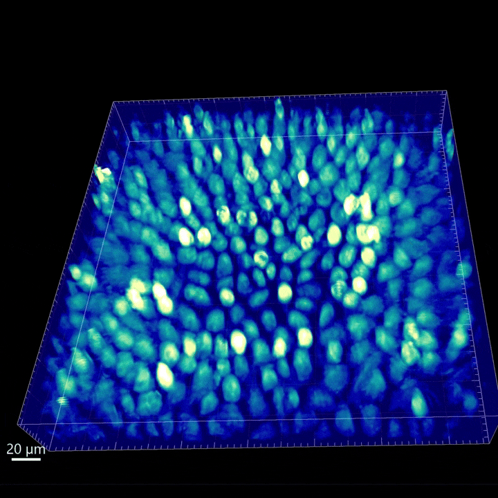

Figure 1:FDT footage obtained with the Kinetix22 on 3D samples. Left is a MDCK cell monolayer (358.4 x 358.4 x 44 µm3), exposure time 20 ms. Right is a muscle tube (530 x 530 x 300 µm3), exposure time 100 ms. Color represents the refractive index.
Challenge
Prof. Xue encounters numerous imaging and optics challenges in her work, here she describes just a few, “While we do use a single layer of MDCK cells to test or validate our techniques, our main samples are too bulky for ODT, some of the thickest ones are cultivated meat muscle tubes over 300 microns thick, and we are definitely aiming to image deeper.”
“For the proof-of-concept experiments, the bulky label-free tissues are cultured in a fluorescence-coated petri dish. Our fluorescent marker emits at around 600 nm so we need a camera sensitive in that wavelength. The diffracted fluorescence light from the thick tissues is very weak, leading to the challenge of signal-to-noise ratio. We hope to image over a big field of view for these large samples, and we also want high resolution imaging, but this will add more burden to our reconstruction algorithm.”
Prof. Xue needs a versatile, powerful imaging solution that can perform ultra-high sensitivity imaging across a large area.
The Kinetix22 is versatile, it can do ultra-fast and ultra-sensitive imaging, this is great for my lab as we want to use a camera for many different projects, I have no complaints with the Kinetix.
Prof. Yi Xue
Solution
Prof. Xue uses the Kinetix22, the ideal solution for this novel application. “We needed the ultra-sensitivity of the Kinetix22 for our fluorescence diffraction tomography experiments. The Kinetix22 is so versatile, it has many different modes and we use the Sub-Electron or Sensitivity modes. This is very sensitive, the exposure time can be relatively long as we don’t need a high frame rate, but it’s doing very well with imaging the weak diffracted fluorescence signal. We have enough signal to noise ratio to reconstruct good images and have completed several manuscripts from this.”
“Set up in micro-manager was very easy, I’ve used a Prime 95B from [Teledyne] Photometrics for many years so I didn’t have any trouble getting the Kinetix22 set up.”
Reference
Renzi H., Yucheng L., Junjie C., Xue Y., (2024) Fluorescence Diffraction Tomography using Explicit Neural Fields, arXiv:2407.16657 [physics.optics],
https://doi.org/10.48550/arXiv.2407.16657