SCAPE Microscopy
Introduction
Advanced imaging applications strive to image biological processes faster, across a larger area, and in three dimensions. Traditional microscopy methods continue to improve but many struggle to keep up with the dynamic and complex nature of live biological systems.
Microscopy has steadily evolved to overcome the limits of speed, depth, and phototoxicity:
• Laser scanning confocal microscopy (1980s): Enabled optical sectioning through point-by-point scanning with a laser. Precise but slow and prone to photobleaching.
• Spinning disk confocal (1990s): Increased speed by parallelizing point scanning via a spinning disk full of pinholes. Ideal for live imaging but had limited imaging depth.
• Light sheet microscopy (2000s): Delivered plane illumination from the side, dramatically reducing photodamage and increasing imaging speed. However, dual-objective geometry made it challenging for many biological specimens
To this end, in 2015 Prof. Elizabeth Hillman and team developed swept confocally-aligned planar excitation microscopy, or SCAPE (Bouchard et al. 2015). SCAPE is a single-objective light-sheet imaging method designed for high-speed volumetric 3D imaging, without moving the objective or sample. A sweeping light sheet synchronized with fast camera acquisition offered true volumetric imaging at unprecedented speeds.
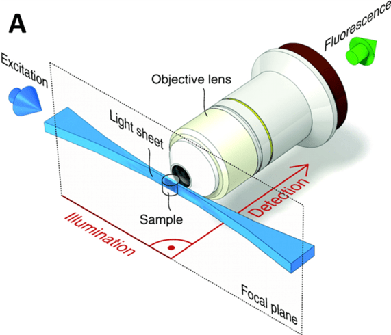
Figure 1: A light sheet microscopy imaging setup. The sample (shown by the clear barrel in image) is illuminated
with a light sheet from the side, exciting fluorophores close to the focal plane. The light sheet is hourglass-shaped
and the thinnest section is used to illuminate the sample. At 90° to this illumination is the objective lens,
which sends emitted fluorescence to the detector camera.
SCAPE Microscopy
SCAPE essentially combines the theory behind confocal and light-sheet microscopy, combining the optical sectioning capabilities of confocal imaging with the orthogonal planar illumination of light-sheet. SCAPE uses the oblique, single-objective format of light-sheet, where the light sheet is rapidly scanned across the sample, resulting in high-speed 3D imaging without needing to move the objective lens or the sample itself, just the illumination.
SCAPE therefore allows for greater freedom with sample geometries, able to image unrestrained or freely moving organisms unlike some formats of light-sheet. However, it is essential to pair SCAPE with a high speed CMOS camera capable of reading out the sample excitation by synchronizing with the scanning light-sheet.
Key advantages of SCAPE include high speed volumetric imaging (10-300 volumes per second), low photodamage to the sample due to the moving light sheet illumination, compatibility with live samples for in vivo imaging, a compact design that can fit onto standard microscope configurations, and SCAPE can be applied to a wide range of different applications, from neuroscience to live cell imaging and whole organism imaging.
SCAPE 2.0
SCAPE microscopy was further iterated upon with SCAPE 2.0, introduced in 2019 (Voleti et al. 2019) and featuring a number of changes and improvements, as seen in Table 1.
|
Feature |
SCAPE (2015) |
SCAPE 2.0 (2019) |
|
Optical Design |
Original layout with more alignment sensitivity |
Improved alignment tolerance and optical stability |
|
Field of View & Resolution |
~400 µm³ volume |
Up to ~1 mm³ volume with cellular resolution |
|
Volumetric Imaging Speed |
~20–100 volumes per second |
Up to ~300 volumes per second |
|
System Footprint |
Custom but somewhat bulky |
More compact, modular, easier to integrate |
|
Aberration Correction |
Limited |
Improved optical correction and image uniformity |
|
Versatility |
Suited for specific demo systems |
More adaptable to different model organisms (e.g., mouse cortex, fly, zebrafish) |
|
Ease of Use |
High-end lab build |
More robust and reproducible, aimed toward broader adoption |
SCAPE 2.0 introduces a Powell lens for more uniform light-sheet illumination, and a Plössl scan lens to reduce aberrations and improve the numerical aperture. With increased imaging speed, it is even more important to pair any SCAPE 2.0 system with a sufficiently large, sensitive and high-speed CMOS camera.
-
- SCAPE Application Examples
With all the benefits of SCAPE and SCAPE 2.0, there are numerous examples of this technology being implemented in research in order to obtain more biologically relevant high speed, 3D imaging data. Here we have two examples of SCAPE in action, both on live cell samples.
Prof. Alf Honigmann of TU Dresden in Germany uses SCAPE microscopy to image live stem cells and epithelial tissues. These samples are highly sensitive to photobleaching and need to be imaged with a low light regime, ideal for light-sheet microscopy and SCAPE. With gentle illumination and scanning at an angle with the light-sheet, Prof. Honigmann can do high-speed 3D volumetric imaging without compromising on cell viability. The team have paired this imaging system with the Kinetix22 CMOS camera, to find out more: Read Here


Figure 2: Top) 2D cross section time lapse video of MDCK cells. Bot) 3D time lapse video of mES cells. All images acquired by the Kinetix22 sCMOS camera.
Prof. Miao Ping Chien’s team at Erasmus University Medical Centre in the Netherlands are using microscopy to investigate aggressive cancer cell subpopulations. They are using a SCAPE microscopy setup for high‑throughput imaging of 3D tissues samples, aiming to screen thousands of samples in a biologically relevant environment. The team have paired this imaging system with the Kinetix22 CMOS camera, to find out more: Read Here

Figure 3: Patient-derived slice of tissue (~300 µm thick) from a head and neck cancer. The volume consists of 500 images with 300 nm spacing, integration time on the Kinetix22 for a single image was 100 ms. Sample was stained with SPY650 as a live cell DNA stain (with 638 nm excitation). Acquired with the Kinetix22 sCMOS camera.
Cameras for SCAPE
SCAPE and SCAPE 2.0 require a CMOS camera with a large sensor, high imaging speeds, high sensitivity (from maximizing signal gain with high quantum efficiency and minimizing noise) and a large pixel array for high spatial image resolution.
If using SCAPE to perform fast volumetric imaging of live organisms, it is vital to ensure the camera can capture the low fluorescent signals from as wide an area as possible, while also synchronizing the readout of the camera to the scanning light sheet. In order to perform faster imaging with SCAPE 2.0, it is vital to use a camera that can read out each pixel row faster than the galvo mirror can scan, otherwise images may contain artifacts. Fortunately, modern CMOS cameras can now read out at such high speeds (line times in the microsecond scale) that selecting the correct camera should eliminate these issues.
Summary
SCAPE microscopy redefines what’s possible in high-speed, 3D biological imaging. Its integration with advanced CMOS cameras unlocks real-time insight into how cells, tissues, and organisms behave in their natural state. As researchers continue to push the boundaries of observation, SCAPE offers a platform that evolves alongside their questions.
References
Bouchard, M. B., Voleti, V., Mendes, C. S., Lacefield, C., Grueber, W. B., Mann, R. S., Bruno, R. M., & Hillman, E. M. (2015). Swept confocally-aligned planar excitation (SCAPE) microscopy for high speed volumetric imaging of behaving organisms. Nature photonics, 9(2), 113–119. https://doi.org/10.1038/nphoton.2014.323
Voleti, V., Patel, K. B., Li, W., Perez Campos, C., Bharadwaj, S., Yu, H., Ford, C., Casper, M. J., Yan, R. W., Liang, W., Wen, C., Kimura, K. D., Targoff, K. L., & Hillman, E. M. C. (2019). Real-time volumetric microscopy of in vivo dynamics and large-scale samples with SCAPE 2.0. Nature methods, 16(10), 1054–1062. https://doi.org/10.1038/s41592-019-0579-4
Further Reading
Back To Learning Center
Contact Us
The Kinetix Family for SCAPE Microscopy