mesoSPIM Volume Imaging
Dr. Tchern Lenn
Microscopy and Imaging Translational Technology Platform, University College London Cancer Institute, UK
Background
Dr. Tchern Lenn is a researcher at the UCL’s Cancer Institute. To understand cancer cell activity and proliferation, it is beneficial to observe their 3D distribution in cancerous tissue/organs using advanced imaging systems, typically light-sheet microscopy due to its suitability for fluorescence imaging of large samples. Dr. Lenn chose the benchtop mesoSPIM design (The mesoSPIM Initiative) and customized it for his institute’s research, mesoSPIM design is highly flexible and affordable without compromising on image quality. The open-source concept allows simple access to anyone willing to acquire the hardware and build the setup.
Dr. Lenn states, “We made a couple of modifications to the electronics and components to make imaging 5-6 channels less expensive, my modifications can be found on the mesoSPIM GitHub repository”, an online platform where hardware design, parts list, assembly instructions and software can be shared but also adapted to the specific requirements of a project.
With his setup, Dr. Lenn supports researchers of the UCL Cancer Institute, imaging a wide range of samples such as in vivo brain cancer in mouse models or in vitro synthetic scaffolds for studying bone cancer. A video sequence demonstrating the type of data generated with the custom mesoSPIM system is displayed in Fig. 1. A cleared mouse brain was rendered in 3D, resolving three different structures. The design of the custom mesoSPIM system leverages high-speed volume imaging by providing an affordable high-quality bespoke design, while promoting a community spirit, summing up the efforts of different researchers into an adaptable framework.
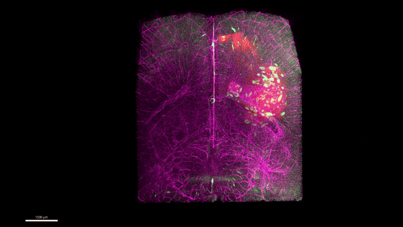
Figure 1: Video of a cleared mouse brain imaged with the modified mesoSPIM based setup using the Iris 15 sCMOS camera. The brain’s vascular system is displayed in magenta, microglia in green and tumour cells in red.
Challenge
Light-sheet microscopy requires accurate triggering of the illumination to synchronize sample excitation with the acquisition, which is paramount to maximise imaging speed and keep the system high-throughput when dealing with large or dynamic biological samples.
This high-throughput imaging also reduces the integration time allocated to every single frame, thus reducing the photon budget for the detection. The camera used with this custom mesoSPIM system therefore must be sensitive enough to account for the limited photon budget. In addition, high resolution imaging is vital to ensure specific sample details can be clearly seen for analysis, requiring a camera that has both a large field of view (FOV) to image large mesoSPIM samples, but also a small enough pixel to achieve sub-cellular resolution across these samples. Furthermore, the open-source concept of the mesoSPIM system requires available SDKs and software wrappers matching the programming language used for controlling the other hardware pieces.
The [Iris 15] camera worked very nicely out-of-the-box... The installation was very easy, and the sensitivity is great for us.
Dr. Tchern Lenn
Solution
The Iris 15 sCMOS camera is an established solution for high quality imaging in a multitude of mesoSPIM systems. In contrast to other camera solutions, the Iris 15 has a large rectangular sensor instead of a squared one, measuring over 25 mm, well suited for imaging large samples. The rectangular sensor is also advantageous for light-sheet microscopy because the larger horizontal dimension better fits the sheet-like illumination and makes imaging of elongated samples such as tissues and organoids more efficient. Combined with the optimised pixel size of 4.25 µm, the Iris 15 is an efficient option for high resolution at different magnifications, specifically designed to excel at light-sheet and mesoSPIM.
The high quantum efficiency and the low noise are features that allow users to reliably image fluorescence even in low light applications, along with user-friendly triggering functionalities freely accessible through a Python API, the Iris 15 is simple to integrate into the existing Python frameworks.
The hardware, the camera and the software implementation leverage this custom high-throughput imaging solution, which is both affordable, powerful, and well-suited to the needs of the UCL Cancer Institute.
![]()