Scanless Two-Photon Voltage Imaging
Dr. Osnath Assayag
Intelligent Imaging Innovations (3i), Paris, France
Background
One of the main interests of the research group at Intelligent Imaging Innovations (3i) is developing and improving different imaging technologies. Recent engineering and conceptual improvements in holographic microscopy hardware have opened the technique to different applications, one feature enabled by holographic microscopy is the possibility to target light in a spatially confined part of a sample using phase-only laser modulation.
Traditional two-photon imaging methods typically rely on point scanning within the sample. While the method is established, the scanning procedure is time-consuming and struggles to capture fast neural dynamics as the imaging speed is intrinsically limited by the mirror speed (less than 100 Hz in Resonant Scanner for a 128 x 128 pixel array). This shortcoming is addressed by scanless two-photon excitation approaches, where holography can target a specific region in the sample and increase the temporal resolution of two-photon imaging by eliminating the delays introduced by mechanical scanning. Scanless two-photon photostimulation is a gateway to collect electrophysiological data, when used in combination with the appropriate hardware and the right sample preparation strategy.
Patch-clamp electrophysiology is the gold standard to record neural activity, but the technique cannot be used to record electrical activity from multiple cells simultaneously and is usually limited to two cells. Its invasive character makes it challenging to perform in vivo and requires highly skilled personnel. Unlike electrophysiology, this approach presented by 3i has a low degree of invasiveness, allowing the simultaneous recording of several neuronal cells and subcellular structures. By engineering the imaging approach to match the photophysical properties of genetically encoded voltage indicators (GEVIs), two-photon voltage imaging can be achieved [1]. This powerful tool overcomes the drawbacks of traditional methods and can be used for a plethora of applications such as high fidelity recording of high frequency spike trains and depolarizations, in vivo imaging of multiple cells up to 250 µm deep in mice brains, simultaneous triggering and imaging of action potentials with one laser source, and much more.
The overall aim of this innovative method is to perform more detailed studies of neural activity, potentially leading to a better understanding of neurological disorders.
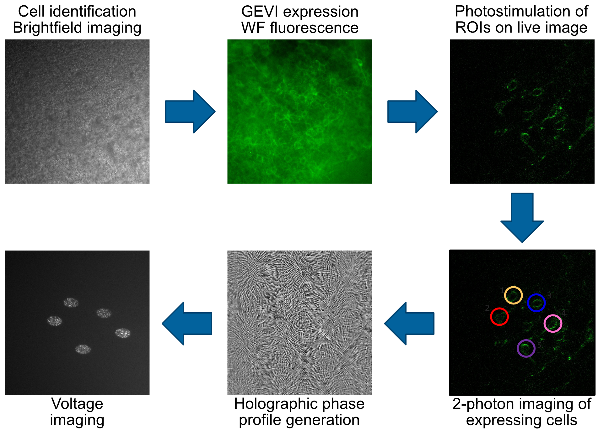
Figure 1: Process of imaging neural activity with voltage sensitive dyes and computer-generated holography.

Figure 2: Top: Jedi-2p-Kv expressing acute slices of the paragigantocellular reticular nucleus [Samples provided by Rémi Fournel and Gilles Fortin ENS, Paris]; Bottom: Scanless two photon voltage imaging performed [2P voltage imaging done in collaboration with R. Sims, Dimitri Tanese, Soledad Dominguez, Imane Bendifallah and Valentina Emiliani (institut de la Vision, Paris)]
Challenge
To record two-photon voltage images with short acquisition times and a high signal to noise ratio (SNR), there are considerable requirements for the detector. To match the temporal sensitivity needed for the experiment, a detector with the capability of collecting data with acquisition frequencies in the range of kHz is paramount. This requires millisecond exposure times, limiting the signal level. In order to image at a high speed, the ideal detector would need to be highly sensitive in order to capture sufficient signal within short exposure windows. In addition, voltage imaging not only involves low signal intensities, but small fluctuations in signal, meaning that sensitivity is paramount in order to capture relevent physiological data.
Both the speed and sensitivity of the detector need to match the application for a scalness two-photon voltage imaging system to function optimally and avoid any imaging compromises.
We are happy with the speed of the Kinetix camera for our experiments.
Dr. Osnath Assayag
Solution
The Kinetix22 sCMOS camera is a versatile and powerful imaging solution, combining the sensitivity, the acquisition flexibility, and the temporal resolution to perform two-photon voltage imaging. In the 8-bit speed mode, a frame rate of >660 fps (Hz) can be achieved across the entire 2400 x 2400 (>5 MP, 22 mm diagonal) sensor, with high imaging speeds also available in 16-bit modes at 118 fps (Hz). Furthermore, a region of interest (ROI) can be set on the chip, reducing the number of pixels to read out and consequently speeding up the image acquisition drastically. When imaging a smaller ROI in the sensitivity mode, an acquisition frequency of >1 kHz is still possible. The quantum efficiency of the Kinetix22 sensor reaches over 95% in the visible part of the spectrum, and with a read noise of less than 1 electron, high quality images can be streamed for voltage imaging. These features establish the Kinetix22 as an optimal detector for scanless two-photon voltage imaging.
Reference
[1] Sims, R.R., Bendifallah, I., Grimm, C. et al. (2024) Scanless two-photon voltage imaging. Nat Commun 15, 5095. https://doi.org/10.1038/s41467-024-49192-2